Biotransformation of two proteratogenic anti-epileptics in the zebrafish (Danio rerio) embryo

02:572 years ago

The zebrafish (Danio rerio) embryo has gained interest as an alternative model for developmental toxicity testing, which still mainly relies on in vivo mammalian models (e.g., rat, rabbit). However, cytochrome P450 (CYP)-mediated drug metabolism, which is critical for the bioactivation of several proteratogens, is still under debate for this model. Therefore, we investigated the potential capacity of zebrafish embryos/larvae to bioactivate two known mammalian proteratogens, carbamazepine (CBZ) and phenytoin (PHE) into their mammalian active metabolites, carbamazepine-10,11-epoxide (E-CBZ) and 5-(4-hydroxyphenyl)-5-phenylhydantoin (HPPH), respectively. Zebrafish embryos were exposed to three concentrations (31.25, 85, and 250 μM) of CBZ and PHE from 51⁄4 to 120 hours post fertilization (hpf) at 28.5°C under a 14/10 hour light/dark cycle. For species comparison, also adult zebrafish, rat, rabbit and human liver microsomes (200 μg/ml) were exposed to 100 μM of CBZ or PHE for 240 minutes at 28.5°C. Potential formation of the mammalian metabolites was assessed in the embryo medium (48, 96, and 120 hpf); pooled (n=20) whole embryos/larvae extracts (24 and 120 hpf); and in the microsomal reaction mixtures (at 5 and 240 minutes) by targeted investigation using a UPLC–Triple Quadrupole MS system with lamotrigine (0.39 μM) as internal standard. Our study showed that zebrafish embryos metabolize CBZ to E-CBZ, but only at the end of organogenesis (from 96 hpf onwards), and no biotransformation of PHE to HPPH occurred. In contrast, our in vitro drug metabolism assay showed that adult zebrafish metabolize both compounds into their active mammalian metabolites. However, significant differences in metabolic rate were observed among the investigated species. These results highlight the importance of including the zebrafish in the in vitro drug metabolism testing battery
for accurate species selection in toxicity studies.

Related

Five simple tricks for making your own video for TPI.tv
Various subjects

Five simple tricks for making your own video for TPI.tv

This video shows you how to make a video yourself. It's really not that difficult! See also https://tpi.tv/how-to-submit for additional information.
01:234 years ago
Stem cell derived Vessels-on-Chip to study brain disorders
Innovation examples
Innovation

Stem cell derived Vessels-on-Chip to study brain disorders

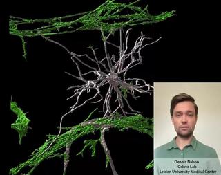
Dennis Nahon is a PhD candidate in the Department of Anatomy and Embryology at the Leiden University Medical Center. In his research, under supervision of Dr. Valeria Orlova (https://www.orlovalab.com/) and Prof. Dr. Christine Mummery, he aims to mimic a blood vessel in the brain by combining different stem cell derived cell types, in a 3D Vessel-on-Chip model. Here, an example of these in vitro blood vessels is shown in which certain brain cells known as astrocytes (in white) interact with the blood vessels (in red). This model paves the way for investigating brain vessels outside the human body, while reducing the need for animal models.
01:532 days ago
 From 2D hiPSC culture to developing a 3D vessel-on-chip
Innovation examples
Innovation

From 2D hiPSC culture to developing a 3D vessel-on-chip

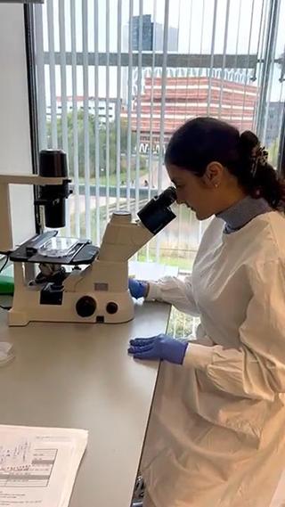
Theano Tsikari is a 2nd year PhD student at the Orlova group at LUMC. As part of the LymphChip consortium, her project focuses on the development of immunocompetent organ-on-chip models of the cardiovascular system, and especially the integration of tissue-resident macrophages and lymphatic vasculature using human induced pluripotent stem cells. In this video, you can follow her as she presents you the backbone of her project, a 3D hiPSC-derived vessel-on-chip model, that has been previously developed in the Orlova group and can be employed for the generation of advanced in vitro models of vascular diseases.
01:292 days ago
Unified organoid system for modeling heart and kidney interaction on-a-chip
Innovation examples
Innovation

Unified organoid system for modeling heart and kidney interaction on-a-chip

Beatrice Gabbin is a PhD candidate at the Anatomy and Embryology Department of the Leiden University Medical Center. Her project is shared with the Nephrology Department and focusses on the study of the cardiorenal axis in vitro. Both heart and kidneys have vital functions in the human body and reciprocally influence each other’s behavior: pathological changes in one can damage the other. There are already multiple independent in vitro (human) models of heart and kidney, but none have so far captured their dynamic crosstalk. The aim of the project is therefore to develop a microfluidic system which can be used to study heart and kidney interaction in vitro. For this purpose, cardiac microtissues and kidney organoids derived from human induced pluripotent stem cells are generated and loaded onto a 3D perfusion chip for their dynamic co-culture. This system enables the study the cardiac and kidney interaction with a high level of control. The validation of a unified organoid system will enable the investigation of diseases involving the two organs and their potential treatments. Read more via the link in the video and https://doi.org/10.1016/j.mtbio.2023.100818.
01:363 days ago