Optimizing CAR-T-cell therapy using 3D tumor models and real-time cell imaging
Chimeric antigen receptor (CAR) T-cell therapy accounts for one of the most promising therapeutic advances in cancer immunotherapy. In this form of adoptive cell transfer, T-cells of a patient are engineered to express so-called ‘CARs’, in which the antigen-recognition capacity of antibodies is combined with T-cell activating domains. So far, CAR-T-cell therapy obtained its most impressive results in hematological malignancies resulting in the approval of five CAR-T cell products by the FDA for hematologic indications. However, CAR-T-cell therapy has not mirrored its success in solid tumors. The poor efficacy of CAR-T-cell therapy in solid tumors has, in part, been attributed to the lack of understanding in how CAR-T-cells function in a solid tumor microenvironment. Classical validation methods rely on the use of specificity and functionality assays in 2D models against adherent target cells or target cells in suspension. Yet, by using these models, observations made in vitro may differ greatly to an in vivo situation where tumors are engrafted in 3D structures. We developed a more relevant and translational 3D tumor model using eGFP+ target cells. This allows us to couple 3D tumor cell killing by CAR-T-cells to live-cell imaging, providing an efficient quantification of target cell death. As proof- of-concept, we used a 3D model of eGFP+ glioblastoma cells and CAR-T-cells targeting a pan-cancer antigen. This 3D glioblastoma model allowed us to show that classical scFv-based CAR-T-cell therapy of glioblastoma cells can be improved by nanoCAR-T-cells. Furthermore, combining nanoCAR-T-cell therapy with a genetic approach of nanobody-based anti-PD-L1 immune checkpoint blockade further increased the cytotoxicity of the nanoCAR-T-cell therapy.
Related
New

Five simple tricks for making your own video for TPI.tv
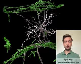
Stem cell derived Vessels-on-Chip to study brain disorders
From 2D hiPSC culture to developing a 3D vessel-on-chip