Popular
Innovation examples
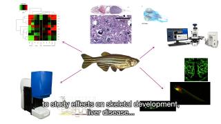
HealthToxicology
Zebrafish in toxicity testing
Zebrafish are increasingly recognised as a useful model for toxicity testing of chemical substances. Testing strategies are becoming more based on mechanisms of toxicity structured in adverse outcome pathways describing the chain of events leading to toxicity or disease. Using a battery of dedicated in vitro and in silico assays, insight can be gained in how exposure leads to disease. For certain diseases it is known that toxicity relies on the interaction between different organs and cell types, which requires research on whole organisms in addition to simple in vitro models. The zebrafish is considered a valuable whole organism model in a mechanism-based testing strategy. At RIVM, the zebrafish embryo model is used for testing the effect of chemical substances on several adverse outcomes and diseases.
For more information see: https://ehp.niehs.nih.gov/doi/10.1289/EHP9888; https://doi.org/10.3390/ijerph18136717; www.linkedin.com/in/harm-heusinkveld
Innovation examples
ToxicologyIn vitroOrgan-on-Chip
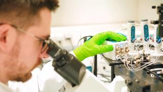
Cartilage-on-a-chip for studying joint degenerative diseases
Carlo Alberto Paggi is currently a PhD candidate at the University of Twente in the research group of Prof. Marcel Karperien and Prof. Séverine Le Gac. Karperien’s lab focus on the biological aspects of osteoarthritic research while Le Gac’s specialize in organ-on-chip development. The project of Carlo Alberto is developing a joint-on-chip platform to create a reliable in vitro model to study disease progression in osteo- or rheumatoid arthritis. The model combines different organ-on-chips aimed at replicating each a tissue around the joint such as cartilage, bone and ligaments. This new technology focuses on better reproducing human models and at substituting the use of animal models for drug research. If you want to know something more about the project and the groups, you can follow the link in the video.
Carlo Paggi was nominated for the Hugo van Poelgeest prize for his research on a cartilage-on-a-chip model to study joint degenerative diseases
Karperien’s lab of Developmental Bioengineering: https://www.utwente.nl/en/tnw/dbe/
Le Gac’s lab of Applied Microfluidics for BioEngineering Research: http://www.severinelegac.com/
Linkedin: https://www.linkedin.com/in/carlo-alberto-paggi-76500b135/
Projects and initiatives
HealthToxicologyIn vitroIn silico
VHP4Safety project
The safety testing of chemicals and pharmaceuticals traditionally relies on animal studies. However, these raise ethical concerns and often fail to accurately predict human responses. New scientific developments offer opportunities to build a Virtual Human Platform (VHP) for safety assessment, a platform that enables assessment based solely on human physiology and biology, integrating data from in vitro and in silico models. This video explains how we are developing the VHP through an interdisciplinary approach. Read the paper in the videolink or visit or VHP4Safety (https://vhp4safety.nl/) for more information.
Innovation examples
ToxicologyIn vitroOrgan-on-Chip
Human pluripotent stem cell derived cardiomyocytes for disease modelling and drug discovery
Berend van Meer did his PhD research in the research group of prof. Christine Mummery at the department of Anatomy and Embryology of the Leiden University Medical Center. In this group, human pluripotent stem cell derived (Organ-on-Chip) models are being developed, mostly cardiovascular models. The work of Berend aimed to understand how well these stem cell based cardiac models can predict the effect of (well-known) drug therapies in patients. Importantly, the outcomes of the experiments were compared to very similar measurements in rabbit heart muscle cells. And while animal models predicted less than 70% correctly, the human stem cell based models predicted almost 80% of the expected effects correctly. The research contributes to understanding the relevance of stem cell based models and strengthens the confidence regulators and pharmaceutical companies have in such models as animal alternatives in the drug development pipeline.
Berend van Meer has won the Hugo van Poelgeest prize 2020 for his research on human pluripotent stem cell derived cardiomyocytes for disease modelling and drug discovery.
Christine Mummery's lab on Heart on Chip, Disease modeling and toxicity: https://www.lumc.nl/org/anatomie-embryologie/research/902040935402533/
Innovation examples
HealthIn vitroOrgan-on-Chip
Using skin and mucosa models to replace animal testing
The skin and mucosa are important tissues that differ between species in health and disease. The group of Sue Gibbs works on the development of advanced in vitro models that mimic these two tissues, specialising in immunity models and organ-on-a-chip technologies. They use skin models to study for example melanoma, skin allergies, eczema, burns and healing wounds. Dental models are used for the safety of materials used in dentistry, for example to test the quality of the implant and false tooth when it comes to attaching to the soft tissue. Their ambition is to expand into the field of multi-organ technology to make even more relevant models for the human skin and mucosa.
Click on the link in the video to watch more or read the interview with Sue he[https://vu.nl/en/research/more-about/using-skin-and-mucosa-models-to-replace-animal-testing]re.
Questions
HelpathonsPolicyBeginner
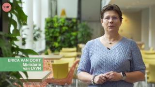
Helpathon #12 – Can you help Erica?
We are inviting Dutch-speaking citizens from all walks of life to join a unique Helpathon and help Erica van Oort, coordinator of the Animal-Free Transition Program (TPI) in the Netherlands. No prior knowledge of animal testing is required—your fresh perspective can help Erica communicate more effectively about animal-free research.
We strongly believe that well-informed citizens are key to improving democratic policy-making on health research, with and without animals. Please share this invitation to at least one suitable person who could contribute—and of course, you are warmly welcome to join as well.

Expert interviews
HealthEducation
Daniela Salvatori, TPI Utrecht: We aim for better science with less animals
Prof. dr. Daniela Salvatori, chair of TPI Utrecht, presents the aims of the local TPI group and invites all who want to share their ideas or questions on the transition towards animal-free innovations to get in touch via uu.nl/tpi.
Meetings & conferences
HealthIn vitroAdvanced
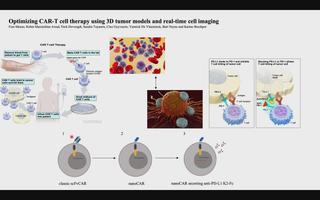
3D tumor models for CAR-T-cell therapy optimization
Chimeric antigen receptor (CAR) T-cell therapy accounts for one of the most promising therapeutic advances in cancer immunotherapy. In this form of adoptive cell transfer, T-cells of a patient are engineered to express so-called ‘CARs’, in which the antigen-recognition capacity of antibodies is combined with T-cell activating domains. So far, CAR-T-cell therapy obtained its most impressive results in hematological malignancies resulting in the approval of five CAR-T cell products by the FDA for hematologic indications. However, CAR-T-cell therapy has not mirrored its success in solid tumors. The poor efficacy of CAR-T-cell therapy in solid tumors has, in part, been attributed to the lack of understanding in how CAR-T-cells function in a solid tumor microenvironment. Classical validation methods rely on the use of specificity and functionality assays in 2D models against adherent target cells or target cells in suspension. Yet, by using these models, observations made in vitro may differ greatly to an in vivo situation where tumors are engrafted in 3D structures. We developed a more relevant and translational 3D tumor model using eGFP+ target cells. This allows us to couple 3D tumor cell killing by CAR-T-cells to live-cell imaging, providing an efficient quantification of target cell death. As proof- of-concept, we used a 3D model of eGFP+ glioblastoma cells and CAR-T-cells targeting a pan-cancer antigen. This 3D glioblastoma model allowed us to show that classical scFv-based CAR-T-cell therapy of glioblastoma cells can be improved by nanoCAR-T-cells. Furthermore, combining nanoCAR-T-cell therapy with a genetic approach of nanobody-based anti-PD-L1 immune checkpoint blockade further increased the cytotoxicity of the nanoCAR-T-cell therapy.
Innovation examples
In vitroOrgan-on-Chip
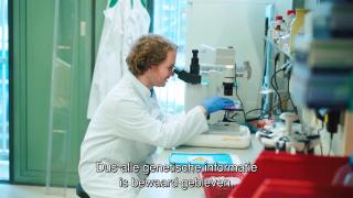
Unified organoid system for modeling heart and kidney interaction on-a-chip
Beatrice Gabbin is a PhD candidate at the Anatomy and Embryology Department of the Leiden University Medical Center. Her project is shared with the Nephrology Department and focusses on the study of the cardiorenal axis in vitro. Both heart and kidneys have vital functions in the human body and reciprocally influence each other’s behavior: pathological changes in one can damage the other. There are already multiple independent in vitro (human) models of heart and kidney, but none have so far captured their dynamic crosstalk. The aim of the project is therefore to develop a microfluidic system which can be used to study heart and kidney interaction in vitro. For this purpose, cardiac microtissues and kidney organoids derived from human induced pluripotent stem cells are generated and loaded onto a 3D perfusion chip for their dynamic co-culture. This system enables the study the cardiac and kidney interaction with a high level of control. The validation of a unified organoid system will enable the investigation of diseases involving the two organs and their potential treatments. Read more via the link in the video and https://doi.org/10.1016/j.mtbio.2023.100818.
Projects and initiatives
HealthToxicology
The NAM Navigator: A unique repository for information on the validation and acceptance of New Approach Methodologies
The NAM navigator is an innovative knowledge portal to navigate you to and through valuable information on the development, standardization, validation and acceptance of New Approach Methodologies (NAM). The NAM Navigator acts as an online guide that provides specific information needed in each of these steps, thereby increasing the broad use of animal-free innovations. Follow the link in the video to start navigating!
Expert interviews
Policy
Charlotte Blattner, Harvard Law School: Transition needs community efforts
Charlotte Blattner is a visiting researcher at Harvard Law School on the Animal Law & Policy Program and explains that a transition is needed to move away from animal testing. This transition needs to be a just transition, a community effort where all stakeholders are involved to replace animal testing for animal-free innovations.
Expert interviews
HealthToxicologyIn vitro
Erwin Roggen, ToxGen Solutions: Applying animal free testing approaches
Erwin Roggen explains his role as pioneer in the development of technologies for animal-free application. His product, ToxGenSolutions, provides test methods required for modern testing and assessment of compounds and products. It builds on a virtual generic platform of leading test and technology developers providing novel technologies addressing key events in adverse outcome pathways.